

Guide to Wrist Anatomy
Wrist Anatomy: The wrist is among the most complex joints in the body. It may be argued that there is no such thing as a single “wrist joint,” since the wrist contains many joints that function together to move the hand.
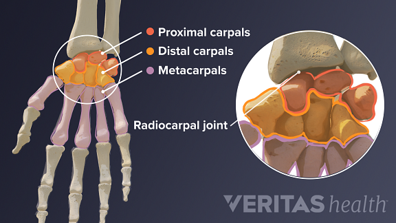
The carpal bones are divided into a proximal row and a distal row. The proximal carpals meet the radius bone at the radiocarpal joint.
The wrist’s functions include:
- Moving the hand back and forth and side to side
- Transferring forces from the arm to the hand
- Providing strength and flexibility to the hand
These functions depend on a complex structure of multiple bones, joints, and soft tissues—tendons, ligaments, nerves, and blood vessels.
Wrist Bones
The bones in and around the wrist consist of the forearm bones, carpal bones, and hand bones.
There are two long bones in the forearm that run from the elbow to the wrist:
- The larger bone, the radius, is on the same side as the thumb.
- The smaller bone, the ulna, is on the little finger side.
The end of the ulna is covered by a triangular-shaped articular disc—a piece of fibrous cartilage that cushions the wrist bones. However, the ulna bone does not directly form a joint with the wrist bones.
Where the radius bone meets the wrist are two rows of small round bones—four in each row—known as carpal bones:
Related Posts
- Proximal carpals: The row of carpal bones closest to the forearm
- Distal carpals: The row of carpal bones closer to the fingers
Together, the eight carpal bones are called the carpus. The distal carpals form five joints with the hand bones. The hand bones are called metacarpal bones. Metacarpals are long bones that connect the distal carpals to the fingers and thumb.
Joints and Motions of the Wrist
There are several sets of joints in and around the wrist. These joints vary in type and have different motions.
- The distal radioulnar joint is located between the radius and the ulna at the wrist. This joint allows for rotation of the forearm. The ulna stays in a stable position while the radius rotates around it.
- The radiocarpal joint is located where the radius meets the first row of carpal bones. This joint is the main joint of the wrist. The radiocarpal joint is condyloid. A condyloid joint allows combined motions in multiple planes, including backward and forward bending motions, side-to-side motions, and circular motions.
- The Midcarpal joint is located where the proximal and distal carpals meet. This joint has features of both condyloid and gliding joints. Gliding joints allow the bones to glide up and down, left and right, and diagonally. The Midcarpal joint performs up-and-down and side-to-side movements and works together with the radiocarpal joint to move the wrist.1
- The carpometacarpal joints are the five joints between the distal carpals and the metacarpals.
- The carpometacarpal joint of the thumb is a saddle joint. This saddle joint allows the thumb to function like a joystick, with forwarding, backward, and side-to-side motions.
- The carpometacarpal joints of the fingers are gliding joints and can perform up-and-down and side-to-side movements. In general, the carpometacarpal joint of the little finger has a greater range of motion compared to the others.
Most wrist fractures are the result of a break in the radius bone at the radiocarpal joint—known as a distal radius fracture. The scaphoid is the second-most commonly fractured wrist bone.
Common wrist motions
The most common wrist movement in daily activities is called the dart thrower’s arc of wrist motion. This action involves bending the wrist backward and toward the thumb, and then forward and toward the little finger. Common everyday activities that involve this motion are hammering a nail, throwing a ball, drinking from a glass, pouring from a jug, and closing the lid of a jar.
On average, the wrist bends at an angle of 30 to 35 degrees backward during extension and 5 to 10 degrees forward during flexion in everyday wrist movements.
The wrist also contains several soft tissues, such as ligaments, tendons, nerves, and blood vessels. These soft tissues function with the wrist’s bones and joints to provide movement, sensation, and nourishment to the hand.
- 1. Kaufmann R, et al. Kinematics of the Midcarpal and radiocarpal joints in radioulnar deviation: an in vivo study. J Hand Surg Am. 2015;30(5):937-42.
- 2. Lee, SK. Fractures of the carpal bones. Green’s Operative Hand Surgery, 7th Ed. Ed. Scott Wolfe. Philadelphia: Elsevier, 2017. 588-652.
- 3. Brigstocke GH, Hearnden A, Holt C, Whatling G. In-vivo confirmation of the use of the dart thrower’s motion during activities of daily living. J Hand Surg Eur Vol. 2014;39(4):373-8.
- 4. Laulan J, Marteau E, Bacle G. Wrist osteoarthritis. Orthopaedics & Traumatology: Surgery & Research. 2015;101(1): S1-S9. doi:10.1016/j.otsr.2014.06.025
Comments are closed.